Vein Ablation

Leg vein ablation is a medical procedure used to treat varicose veins, which are enlarged veins that appear twisted or bulging under the skin. This condition can cause discomfort, pain, and swelling, and can also be unsightly.

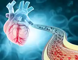
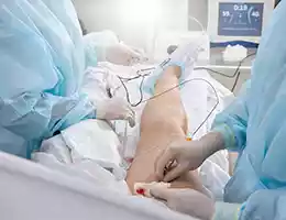
During a leg vein ablation procedure, a small incision is made near the affected vein, and a thin catheter is inserted into the vein. The catheter then delivers energy, such as laser or radiofrequency energy, to the inside of the vein, causing it to heat up and seal shut. This redirects blood flow to healthier veins, improving circulation and reducing the appearance and symptoms of varicose veins.

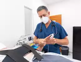
Leg vein ablation is a minimally invasive procedure that is typically done on an outpatient basis, meaning patients can return home the same day. Local anesthesia is used to numb the area, and sedation may be given to help patients relax during the procedure.

After the procedure, patients may need to wear compression stockings for a few weeks to help reduce swelling and promote healing. Normal activities can usually be resumed within a few days, but heavy lifting and strenuous exercise should be avoided for several weeks.

Leg vein ablation is a safe and effective treatment option for varicose veins, and can improve quality of life for those who suffer from this condition. However, as with any medical procedure, it is important to discuss the risks and benefits with a qualified healthcare provider before making a decision.

Cardiology Testing

A cardiology office is a medical practice that specializes in the diagnosis, treatment, and management of heart-related conditions. Some of the tests that may be performed in a cardiology office include:

  • Electrocardiogram (ECG or EKG): An ECG is a non-invasive test that records the electrical activity of the heart. This test can help diagnose heart rhythm disorders, such as atrial fibrillation or ventricular tachycardia.
  • Echocardiogram: An echocardiogram is a non-invasive test that uses ultrasound technology to create images of the heart. This test can help diagnose structural heart problems, such as heart valve disorders or congenital heart defects.
  • Stress tests: Stress tests are medical tests that are used to evaluate the function of the heart during physical activity. These tests can help diagnose heart disease and evaluate the effectiveness of treatment plans.
  • Holter monitor: A Holter monitor is a small, wearable device that records the electrical activity of the heart over a period of time, usually 24 to 48 hours. This test can help diagnose heart rhythm disorders that may not be detected during an ECG.
  • Cardiac catheterization: Cardiac catheterization is an invasive test that involves inserting a thin, flexible tube (catheter) into a blood vessel in the arm or leg and threading it up to the heart. This test can help diagnose blockages in the coronary arteries and other heart-related conditions.
  • CT scans and MRIs: CT scans and MRIs are imaging tests that can help diagnose heart disease and evaluate the effectiveness of treatment plans.

In addition to these tests, a cardiology office may also perform diagnostic procedures such as angiograms, electrophysiology studies, and cardiac ablation therapy.

If you are experiencing symptoms of heart disease, such as chest pain, shortness of breath, or palpitations, it is important to seek medical attention from a qualified healthcare provider. A cardiology office can provide specialized care and diagnostic testing to help diagnose and treat heart-related conditions.

Echocardiograms

An echocardiogram is a diagnostic test that uses ultrasound waves to create images of the heart. It is a non-invasive test that is painless and usually takes less than an hour to complete. Echocardiograms are commonly used to diagnose and monitor a variety of heart conditions, including heart disease, heart valve disorders, and congenital heart defects. In this blog post, we will discuss what an echocardiogram is, how it works, and its uses and limitations.

What is an echocardiogram?

An echocardiogram is a test that uses high-frequency sound waves (ultrasound) to create images of the heart. The test is painless and does not involve any radiation. During the test, a trained technician will place a small device called a transducer on the chest, which emits sound waves that bounce off the heart and create images on a monitor. The test usually takes less than an hour to complete and is typically done on an outpatient basis.

How does an echocardiogram work?

An echocardiogram works by using ultrasound waves to create images of the heart’s structure and function. The transducer emits sound waves that bounce off the heart’s structures, creating images that are displayed on a monitor. The images show the heart’s chambers, valves, and blood flow, allowing doctors to see how the heart is working and diagnose any abnormalities.

There are several types of echocardiograms, including:

  1. Transthoracic echocardiogram (TTE): This is the most common type of echocardiogram. It is done by placing the transducer on the chest and moving it around to get different views of the heart.
  2. Stress echocardiogram: This type of echocardiogram is done before and after exercise to evaluate the heart’s function during physical activity.
  3. Transesophageal echocardiogram (TEE): This type of echocardiogram is done by placing the transducer down the throat and into the esophagus to get a closer look at the heart.

Uses of echocardiograms

Echocardiograms are used to diagnose and monitor a variety of heart conditions, including:

  1. Heart disease: Echocardiograms can help diagnose and monitor heart disease, including heart failure and coronary artery disease.
  2. Heart valve disorders: Echocardiograms can help diagnose and monitor heart valve disorders, including mitral valve prolapse, aortic stenosis, and regurgitation.
  3. Congenital heart defects: Echocardiograms can help diagnose and monitor congenital heart defects, including atrial septal defects and ventricular septal defects.
  4. Cardiomyopathy: Echocardiograms can help diagnose and monitor cardiomyopathy, a condition in which the heart muscle becomes enlarged or thickened.

Limitations of echocardiograms

While echocardiograms are a valuable diagnostic tool, they do have some limitations. Echocardiograms cannot detect all types of heart conditions, and abnormal echocardiograms do not always indicate a serious problem. Other tests, such as cardiac catheterization, may be needed to confirm a diagnosis.

Additionally, echocardiograms are operator-dependent, meaning that the quality of the images depends on the technician’s skill and experience. In some cases, the images may be difficult to interpret, which can lead to misdiagnosis or delayed treatment.

Conclusion

An echocardiogram is a non-invasive, painless diagnostic test that uses ultrasound waves to create images of the heart. It is a valuable tool for diagnosing and monitoring a variety of heart conditions, including heart disease, heart valve disorders, and congenital heart defects. Patients should discuss the benefits and limitations of echocardiograms with their health care provider.

TAVR

Transcatheter Aortic Valve Replacement (TAVR) is a minimally invasive procedure used to treat aortic valve stenosis, a condition in which the aortic valve becomes narrowed and obstructs blood flow from the heart to the rest of the body. TAVR is an alternative to traditional open-heart surgery for patients who are considered high-risk or too frail for surgery.

During a TAVR procedure, a thin, flexible tube called a catheter is inserted through an artery in the groin or chest and guided to the heart. A new valve is then placed over the existing valve using the catheter. Once the new valve is in place, the catheter is removed and the incision is closed.

One of the major benefits of TAVR is its minimally invasive nature. Unlike traditional open-heart surgery, TAVR does not require the breastbone to be cut and the chest to be opened. This results in less pain and a shorter recovery time for patients.

TAVR has been shown to be an effective treatment for aortic valve stenosis in high-risk patients. It has also been approved for patients who are considered intermediate-risk, expanding the number of patients who may benefit from this procedure.

As with any medical procedure, TAVR does carry some risks. These may include bleeding, infection, stroke, or damage to the blood vessels or heart. However, the risks of TAVR are generally lower than those associated with traditional open-heart surgery.

If you are experiencing symptoms of aortic valve stenosis, such as chest pain, shortness of breath, or fatigue, talk to your doctor about whether TAVR may be a suitable treatment option for you. Your doctor can help you weigh the benefits and risks of TAVR and determine the best course of treatment for your individual needs.

Stress Tests

Stress tests are medical tests that are used to evaluate the function of the heart and to diagnose heart-related conditions. These tests are often used in people who have symptoms of heart disease, such as chest pain or shortness of breath.

There are several different types of stress tests, including:

  • Exercise stress test: This is the most common type of stress test. During an exercise stress test, you will walk on a treadmill or pedal a stationary bike while your heart rate and blood pressure are monitored. This allows healthcare providers to evaluate the heart’s response to physical activity.
  • Nuclear stress test: During a nuclear stress test, a small amount of radioactive dye is injected into your bloodstream. Special cameras are then used to take images of your heart while you are at rest and after exercising. This allows healthcare providers to evaluate the blood flow to your heart muscle.
  • Stress echocardiogram: During a stress echocardiogram, an ultrasound is used to create images of the heart at rest and after exercising. This allows healthcare providers to evaluate the heart’s function and blood flow.

Stress tests are generally safe and non-invasive, although there is a small risk of complications such as chest pain, dizziness, or irregular heart rhythms. Before undergoing a stress test, your healthcare provider will perform a physical exam and may order additional tests to ensure that you are a good candidate for the test.

If you are undergoing a stress test, there are several things you can do to prepare. You may be asked to avoid caffeine, nicotine, or certain medications before the test. You should also wear comfortable clothing and shoes, and be prepared to exercise at a moderate intensity.

In summary, stress tests are medical tests that are used to evaluate the function of the heart and to diagnose heart-related conditions. There are several different types of stress tests, including exercise stress tests, nuclear stress tests, and stress echocardiograms. If you are undergoing a stress test, it is important to follow your healthcare provider’s instructions and to take steps to prepare for the test. By doing so, you can help ensure an accurate diagnosis and effective treatment plan for any heart-related conditions.

Stents

Coronary stents are small, expandable mesh tubes that are used to treat coronary artery disease (CAD). They are designed to hold open narrowed or blocked coronary arteries and improve blood flow to the heart muscle. In this blog post, we will discuss what coronary stents are, how they work, and their benefits and risks.

What are coronary stents?

Coronary stents are medical devices that are placed inside a coronary artery to hold it open and improve blood flow. They are made of metal (such as stainless steel or cobalt-chromium) or a combination of metal and polymer. Some stents are coated with medication to help prevent the formation of scar tissue inside the artery.

How do coronary stents work?

Coronary stents are placed inside a coronary artery during a procedure called percutaneous coronary intervention (PCI). During the procedure, a thin, flexible tube called a catheter is threaded through a blood vessel in the groin or arm and guided to the blocked artery in the heart. A small balloon at the tip of the catheter is inflated to compress the plaque and widen the artery. Once the artery is widened, the stent is placed inside to keep the artery open.

Benefits of coronary stents

Coronary stents have several benefits for patients with CAD:

  1. Improved blood flow to the heart: By holding open narrowed or blocked coronary arteries, stents improve blood flow to the heart muscle and reduce the risk of chest pain (angina) and heart attack.
  2. Reduced need for repeat procedures: Stents can help prevent the recurrence of blockages in the coronary arteries, reducing the need for repeat procedures such as PCI or coronary artery bypass graft (CABG) surgery.
  3. Shorter hospital stay: Stenting is a minimally invasive procedure that can be performed on an outpatient basis or with a short hospital stay, allowing patients to return to their normal activities sooner.

Risks of coronary stents

While coronary stents have many benefits, there are also some risks associated with the procedure:

  1. Bleeding: There is a risk of bleeding at the insertion site of the catheter.
  2. Infection: There is a small risk of infection at the insertion site or in the bloodstream.
  3. Blood clots: Stents can increase the risk of blood clots forming inside the artery, which can lead to a heart attack or stroke. To reduce this risk, patients may need to take blood-thinning medications such as aspirin or clopidogrel for several months after the procedure.
  4. Restenosis: In some cases, the artery may become narrowed again (restenosis) after stenting. This may require repeat procedures or surgery.
  5. Stent thrombosis: Rarely, a blood clot may form inside the stent, leading to a sudden blockage of the artery and a heart attack.

Conclusion

Coronary stents are a valuable tool for the treatment of coronary artery disease. They can improve blood flow to the heart, reduce the need for repeat procedures, and shorten hospital stays. However, like all medical procedures, there are risks associated with stenting. It is important for patients to discuss the benefits and risks of stenting with their healthcare provider to determine if it is the right treatment option for them.

FAQs

How long is my blood work valid for?

Blood work done prior to any procedure is usually good for no more than 30 days. Blood work in general may be good for up to a year, although this may vary depending on the situation, and at the discretion of your provider.

How long does testing take?

  • Expect an echocardiogram appointment to last 30-45 minutes.
  • Expect an carotid ultrasound appointment to last 30-45 minutes.
  • Expect an arterial/leg ultrasound appointment to last 30-45 minutes.
  • Expect a treadmill stress test appointment to last 30-45 minutes.
  • Expect a nuclear stress test (either treadmill or pharmacological/chemical) appointment to last 3-4 hours.

How long can I expect an appointment to be?

If you are a new patient to a cardiology practice, anticipate a longer initial visit to complete paperwork and oftentimes obtain records from your other medical providers. Also anticipate a longer visit due to your cardiologist gathering your full prior history and performing a thorough initial physical exam. You will likely have an EKG done on your first visit, and all of your medications will be reviewed. Your first visit may take 1-2 hours overall.

A follow up visit may be varied in length, depending on the nature of the visit. Anticipate up to 60 minutes for a follow up visit. This may be even longer depending if you, or even people before you, need a longer visit to ensure all your needs are addressed.

Who are some of the best cardiologists in Las Vegas?

There is no perfect answer to this question. Just like asking for restaurant reviews, the ‘best’ cardiologist for you is the one that you are the most comfortable with, knows your history, is able to diagnose and treat you appropriately. Not every treatment plan is the best for everyone, and you should have a cardiologist that works best with and caters to you. There are quite a few cardiology groups in the Las Vegas area that have established a respected reputation with patients. Las Vegas Heart Associates, Heart Center of Nevada, Nevada Heart and Vascular Center, Nevada Cardiology, Desert Cardiology, and Intermountain Healthcare all have very well respected cardiologists that are well liked and have good outcomes for the Las Vegas community.

How do I get a copy of my medical records?

If you need copies of any visits, testing, or treatment done at the office, just call or ask your cardiologist’s office in person. You always have the right to see your own records. If you want to obtain your records from the hospital, just ask the medical records department at the hospital.

What is a cardiologist?

A cardiologist is a physician that specializes in people’s heart health. This can be varied in its scope, from preventive care to treatment of active conditions in the hospital or office. Preventive cardiology care may include proactively managing blood pressures, cholesterol levels, diabetes, or even just simple lifestyle and diet modifications to prevent the development of cardiovascular disease. Your cardiologist may talk about weight loss and an exercise regimen to prevent or treat disease. They may also talk about what medications work best for you.

A cardiologist has typically gone through medical school, followed by training in internal medicine. This is followed by several years of training in general cardiology, and possibly followed by further specialization in specific areas of cardiology such as electrophysiology, interventional cardiology, heart failure and transplant, or imaging, to name a few.

What are the types of cardiologists?

There are many different areas that a cardiologist may specialize in. A general cardiologist helps to manage a person’s overall cardiovascular health, including blood pressure, lipid, or diabetes management, or people with existing conditions, such as coronary or peripheral arterial disease, arrhythmias, atrial fibrillation, or pacemaker/defibrillator management, and others. Always ask your cardiologist what his or her specific areas of interest are.

What is the difference between a general cardiologist and an interventional cardiologist?

A general (sometimes called diagnostic) cardiologist will help to manage a person’s overall cardiovascular health. This may include treatment of blood pressure, cholesterol, or diabetes, as well as treatment of arrhythmias or coronary artery disease. He or she may perform diagnostic procedures, such as cardiac catheterizations or transesophageal echocardiograms. An interventional cardiologist (interventionalist) may do all of these procedures as well, but can also treat arterial blockages by using balloons or placing stents to keep an artery open. A general cardiologist will typically work together with an interventionalist and the patient to determine the best course of action for the patient’s disease.

What is an electrophysiologist?

An electrophysiologist cardiologist, or EP, is a cardiologist that specializes in heart rhythm management and treatment. These conditions typically include bradycardia (slow heart rates), tachycardia (fast heart rates), atrial fibrillation (AFib or AF), atrial flutter (Aflutter), supraventricular tachycardia (SVT), ventricular tachycardia (VT), and others. An electrophysiologist will see patients in the hospital or office, and will offer various procedures for treatment of a person’s arrhythmias.

What is a cardiac catheterization?

A cardiac catheterization can go by different names, including a cardiac or heart cath or coronary angiogram. This typically involves going through one’s wrist (radial approach) or leg (groin or femoral approach). A tube (sheath) is placed in the artery, and through that, catheters (long plastic tubes) are passed up to the heart over a wire. These catheters are then placed into the arteries of the heart (coronary arteries), and contrast (dye) is injected into the arteries. The cardiologist performing the procedure then looks under XRay for any evidence of blockages in the arteries to the heart. If there are any blockages, a decision can then be made (with the patient’s involvement) on the best way to treat the blockage. At the same time, another sheath may be placed in the vein, with catheters passed up to the heart and lung to measure pressures in both. This may help diagnose certain lung diseases.

What are the risks of an angiogram?

The biggest risk of a cardiac catheterization is typically bleeding from the site of the procedure, and that risk is usually decreased by using a radial approach for the procedure. There is also a small risk for damaging the arteries that the procedure is performed through, or even of the arteries of the heart. There is also a small risk of heart attack, death, or stroke from the procedure. Because these procedures are typically performed in a hospital, in a controlled setting with physicians, nurses, and appropriate staff involved, the risks are greatly reduced. Always ask the physician performing the procedure about the risks and benefits beforehand.

What is a stress test? What are the different types of stress tests?

Your cardiologist may order a stress test to determine your cardiovascular status, and assess your risk of cardiac events.

The most basic type of stress test is a treadmill stress test. For this, you will be asked to walk on a treadmill under supervision. Your vital signs, including blood pressure and heart rate, will be watched as you walk on a treadmill. You will be hooked up to an EKG to look at the electrical function of your heart. The speed and incline of the treadmill will gradually increase over several minutes, and you will be assessed for how long you can walk, as well as how your vital signs and EKG looks. This may give clues on signs of cardiac or coronary artery disease.

If you cannot walk on a treadmill, or your cardiologist decides this is not the best for you, you may be given medicine (Lexiscan) to stimulate the heart, and again look for signs of heart disease or coronary artery disease on your EKG. The side effects of this medicine (Lexiscan or regadenoson) include arrhythmias, chest pain, shortness of breath, palpitations, or extremely rarely heart attack or death. The test is always performed with staff and a cardiologist present, so even the rare risks are minimized.

In addition to monitoring your EKG with either a treadmill or chemical stress test, your cardiologist may want to take pictures of your heart before and after the stress for comparison. One way is an echocardiogram ( stress echocardiogram) to look at ultrasound images of your heart with rest and stress. Another way is by injecting a contrast that uses nuclear activity picked up by a special camera, to look for areas of concern with the heart.

What is an EKG?

An electrocardiogram, sometimes abbreviated as an ECG or EKG, is an electrical reading of your heart. It can tell if there are any active rhythm issues or if you are having a heart attack. It may also tell if there are signs of damage to the heart, or risk of having heart issues in the future. You will have 10 leads attached to your chest and body, for a total of a 10 second reading.

What is a Holter monitor?

A Holter monitor is a long-term electrical reading of your heart. It is similar to an EKG, and can be worn at home for 24 hours or up to 2 weeks. It is used to detect any rhythm issues while it is being worn.

What is a loop recorder?

A loop recorder is a heart monitor that is implanted under the skin. Unlike external EKGs and Holter monitors that are limited in their length of time worn, a loop recorder has up to a four year battery life. As it is under the skin, your activity afterwards is not limited in any way. The loop recorder can help detect heart rhythm issues, such as atrial fibrillation, or slow or fast heart rates. It can helpful in patients with frequent symptoms such as syncope (passing out) and palpitations, as it can help include or exclude underlying arrhythmias.

What can I do after a pacemaker?

After you have your pacemaker implanted, you should avoid getting it wet for a few days after the procedure. For a few weeks after that, you will have to limit use of your arm on the side of the pacemaker, including avoiding lifting your arm above your head, lifting heavy objects, and driving with that arm.

What can I do after bypass surgery?

After bypass surgery, you will receive instructions from the hospital about activities you can and cannot do. For example, you will have to avoid anything that strains your chest wall for several weeks to months. This includes avoiding using your arms to get up from a chair, lifting anything heavy, or doing any swinging motions with your arms.

What is a CardioMems monitor?

A CardioMems monitor is an implantable device that goes into the artery to your lung. It is implanted through a vein, typically in the leg, as a same-day procedure. Once it is implanted, it can help monitor the pressures inside of the lung to detect worsening signs of heart failure. These measurements can help guide your provider to adjust your diuretics (water pills) so that your heart failure symptoms, such as shortness of breath or leg swelling, are greatly reduced in frequency and severity.

What is an Impella?

An Impella is a device that goes through the artery in the leg to help the function of the heart. In an emergency situation, such as a heart attack, cardiac arrest, or acute heart failure and shock, it can help the heart beat stronger and take the workload off the heart. Similarly, when used electively, it can reduce the work of the heart so it pumps more efficiently, such as during stent procedures.

Palpitations

Palpitations are an abnormal awareness of the heartbeat, which can feel like a fluttering or racing sensation in the chest. While palpitations are often harmless, they can also be a sign of an underlying heart condition. If you experience palpitations, it is important to seek medical attention to determine the cause and receive appropriate treatment.

Some of the most common causes of palpitations include:

  • Anxiety and stress: Palpitations can be caused by anxiety or stress, as these conditions can increase the release of adrenaline in the body, which can affect the heart rate.
  • Caffeine and other stimulants: Consuming caffeine, nicotine, or other stimulants can cause palpitations by stimulating the heart.
  • Medications: Some medications, such as beta-blockers or thyroid medication, can cause palpitations as a side effect.
  • Heart rhythm disorders: Palpitations can also be caused by heart rhythm disorders, such as atrial fibrillation or supraventricular tachycardia.
  • Structural heart problems: Rarely, palpitations can be a sign of a structural problem with the heart, such as a heart valve disorder.

If you experience palpitations, it is important to seek medical attention to determine the cause and receive appropriate treatment. Your healthcare provider may perform tests, such as an electrocardiogram (ECG) or echocardiogram, to evaluate the heart’s rhythm and function. Treatment will depend on the underlying cause of the palpitations and may include medications, lifestyle changes, or procedures such as ablation therapy.

In addition to seeking medical attention, there are steps you can take to help prevent palpitations. This includes avoiding triggers such as caffeine or other stimulants, managing stress and anxiety, and maintaining a healthy lifestyle through regular exercise and a balanced diet.

In summary, palpitations are an abnormal awareness of the heartbeat and can have many different causes. If you experience palpitations, seek medical attention to determine the cause and receive appropriate treatment. Taking steps to prevent palpitations, such as avoiding triggers and managing stress, can also help reduce your risk of experiencing this symptom in the future.

Pacemakers

A cardiac pacemaker is a medical device that is used to regulate the heartbeat of individuals who have an abnormal heart rhythm. The device is implanted under the skin of the chest and connected to the heart by wires. The pacemaker sends electrical signals to the heart to regulate the heartbeat, ensuring that the heart beats at a regular and appropriate rate.

There are several types of pacemakers, and each is designed to address different types of heart rhythm disorders. The two most common types of pacemakers are single-chamber pacemakers and dual-chamber pacemakers. Single-chamber pacemakers are used when the problem is with the heart’s lower chambers (ventricles), while dual-chamber pacemakers are used when the problem is with both the upper and lower chambers (atria and ventricles).

The pacemaker is programmed to deliver electrical signals to the heart at a rate and rhythm that is appropriate for the individual patient. The pacemaker’s programming can be adjusted as needed to accommodate changes in the patient’s heart rhythm or overall health status.

In addition to regulating the heartbeat, pacemakers can also monitor the heart’s activity and store information about the heart’s rhythm and function. This information can be downloaded and analyzed by healthcare providers to help diagnose and treat heart rhythm disorders.

While pacemakers are generally safe and effective, they do require regular monitoring and maintenance. Patients with pacemakers must attend regular follow-up appointments with their healthcare providers to ensure that the device is working properly and to make any necessary adjustments to the pacemaker’s programming.

A pacemaker is a small device that helps regulate your heartbeat. It is commonly used in patients who have an abnormal heart rhythm, known as arrhythmia. After a pacemaker implantation, it is important to take good care of yourself to ensure the device functions correctly and you remain healthy.

Here are some tips for caring for yourself after a pacemaker:

  • Follow your doctor’s instructions: Your doctor will provide specific instructions on how to care for your pacemaker, including how to take care of the incision site, how to use your arm on the side of the implant, and what activities to avoid.
  • Keep your incision clean and dry: To prevent infection, keep the area around the pacemaker implant clean and dry. Your doctor will likely provide instructions on how to care for the incision site and what signs to look out for in case of infection.
  • Avoid heavy lifting and strenuous activity: Your doctor may recommend avoiding heavy lifting and strenuous activity for several weeks after the implantation. This is to avoid putting any unnecessary strain on the implant site and allow it to heal properly.
  • Avoid electromagnetic fields: Pacemakers can be affected by electromagnetic fields, which can disrupt the device’s function. Avoid using devices such as microwave ovens and metal detectors, and keep a safe distance from high voltage power lines.
  • Check your pulse regularly: It is important to monitor your heart rate regularly to ensure that the pacemaker is functioning correctly. Your doctor will provide instructions on how to check your pulse and what to look out for if you experience any symptoms.
  • Keep a record of your device: It is important to keep a record of your pacemaker model and serial number in case of emergency situations. This information can help medical personnel quickly identify the device and provide appropriate care.
  • Attend follow-up appointments: Regular follow-up appointments with your doctor are important to ensure that the pacemaker is functioning correctly and to monitor any changes in your condition. Your doctor will provide instructions on how often you should schedule follow-up appointments.

A pacemaker is a small, battery-operated device that helps regulate an individual’s heartbeat. It is implanted under the skin of the chest, and it uses electrical impulses to keep the heart beating at a regular pace. Pacemakers are commonly used to treat heart conditions such as bradycardia, tachycardia, and heart failure. There are several types of pacemakers available, and each is designed to meet the specific needs of the patient.

  • Single-chamber pacemaker: A single-chamber pacemaker is designed to stimulate only one chamber of the heart, either the atrium or the ventricle.
  • Dual-chamber pacemaker: A dual-chamber pacemaker is designed to stimulate both the atrium and the ventricle of the heart. This type of pacemaker can help improve the coordination of the heartbeat between the two chambers.
  • Biventricular pacemaker: A biventricular pacemaker, also known as a cardiac resynchronization therapy (CRT) device, is designed to treat heart failure. This type of pacemaker stimulates both ventricles of the heart at the same time, helping to improve the coordination of the heartbeat and increase the heart’s pumping efficiency.
  • Leadless pacemaker: A leadless pacemaker is a new type of pacemaker that does not require leads, which are the wires that connect the pacemaker to the heart. Instead, the pacemaker is implanted directly into the heart, making it less invasive than traditional pacemakers.
  • Implantable cardioverter-defibrillator (ICD): An ICD is a type of pacemaker that is designed to treat life-threatening arrhythmias, such as ventricular fibrillation. In addition to regulating the heartbeat, an ICD can also deliver an electric shock to the heart if it detects a dangerous arrhythmia.
  • Subcutaneous ICD (S-ICD): An S-ICD is similar to an ICD, but it is implanted under the skin of the chest instead of inside the heart. This type of pacemaker is less invasive than traditional ICDs and does not require leads to be placed inside the heart.

There are several types of pacemakers available, each designed to meet the specific needs of the patient. If you are considering a pacemaker, it is important to discuss the different options with your doctor to determine which type of pacemaker is best for you. With the help of a pacemaker, you can improve your quality of life and reduce the risk of complications associated with heart conditions.

Cardiac pacemakers are an important tool in the management of heart rhythm disorders. They provide a safe and effective way to regulate the heartbeat, improving the quality of life for millions of people around the world. If you or a loved one has a heart rhythm disorder, talk to your healthcare provider about whether a pacemaker may be a suitable treatment option.

In conclusion, caring for yourself after a pacemaker implantation is critical to ensuring that the device functions correctly and you remain healthy. By following your doctor’s instructions, keeping the incision site clean and dry, avoiding heavy lifting and strenuous activity, monitoring your heart rate, avoiding electromagnetic fields, keeping a record of your device, and attending follow-up appointments, you can ensure a smooth recovery and long-term success with your pacemaker.

Heart Attack

A heart attack, also known as a myocardial infarction (MI), occurs when there is a blockage in one or more of the coronary arteries that supply blood to the heart muscle. This blockage prevents the heart muscle from receiving the necessary oxygen and nutrients, leading to damage and potentially irreversible cell death.

Symptoms of a heart attack can vary from person to person, but the most common symptoms include chest pain or discomfort that may radiate to the arm, shoulder, back, or jaw. Other symptoms may include shortness of breath, nausea, vomiting, sweating, and lightheadedness.

Risk factors for heart attacks include smoking, high blood pressure, high cholesterol, diabetes, obesity, family history, and a sedentary lifestyle. It is important to manage these risk factors through lifestyle changes and/or medication to prevent the development of heart disease.

If you suspect that you or someone you know is having a heart attack, it is important to seek immediate medical attention. Treatment for a heart attack may include medication to reduce clotting and restore blood flow, cardiac catheterization to open blocked arteries, and/or surgery to repair or bypass damaged arteries.

Prevention of heart attacks involves a healthy lifestyle, including a balanced diet, regular exercise, not smoking, and managing risk factors such as high blood pressure and cholesterol levels. It is also important to be aware of the signs and symptoms of a heart attack and to seek medical attention immediately if they occur.

After a heart attack, your recovery process will depend on the severity of the heart attack, the treatment you received, and your overall health. Recovery can take several weeks to several months, and you may need to make lifestyle changes to prevent future heart problems.

Here are some general expectations for the recovery process after a heart attack:

  • Hospitalization: You will likely spend a few days in the hospital after a heart attack for monitoring and treatment.
  • Medications: You will be prescribed medications to prevent blood clots, lower blood pressure, reduce cholesterol levels, and manage any other health conditions you may have.
  • Cardiac rehabilitation: Your healthcare provider may recommend a cardiac rehabilitation program that includes supervised exercise, education on lifestyle changes, and emotional support.
  • Lifestyle changes: You may need to make changes to your diet, exercise routine, and overall lifestyle to reduce your risk of future heart problems. This may include quitting smoking, losing weight, reducing stress, and managing any underlying health conditions.
  • Follow-up appointments: You will need to have regular follow-up appointments with your healthcare provider to monitor your recovery and adjust your treatment plan as needed.
  • Emotional support: It is common to experience emotional distress after a heart attack. Your healthcare provider may recommend counseling or support groups to help you cope with the emotional effects of a heart attack.

It is important to follow your healthcare provider’s recommendations for treatment and lifestyle changes to prevent future heart problems. With proper care and management, many people are able to recover from a heart attack and lead healthy, active lives.

In conclusion, a heart attack is a serious and potentially life-threatening condition that requires prompt medical attention. Managing risk factors and leading a healthy lifestyle can help prevent the development of heart disease and reduce the risk of a heart attack. Remember to seek immediate medical attention if you or someone you know is experiencing symptoms of a heart attack.